Acute Otitis Media: Which is the most important sign for diagnosis?

Posted: Friday, November 19, 2010 | Posted by Debajyoti Datta | Labels: , , 7 comments

Continuing the discussion from my previous post, a systematic review of diagnosis and management of Acute Otitis Media (AOM) has been published in the Journal of the American Medical Association. In light of the above, it would be appropriate to undertake a short review of the pathology and the clinical features of acute otitis media.

Pathophysiology
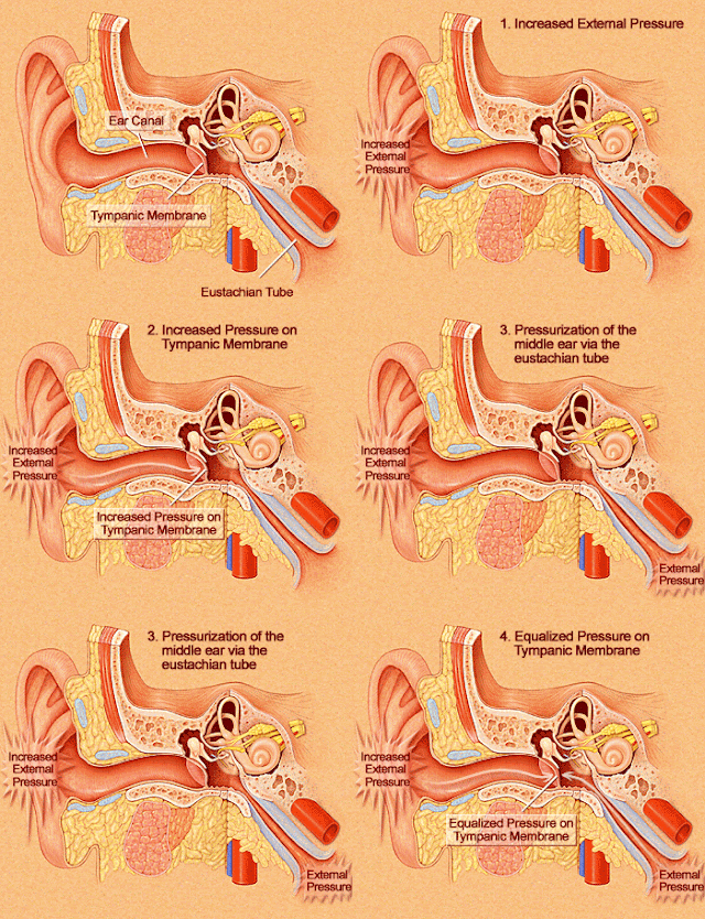
Changes in the middle ear cleft can be described in stages.

pathology and pathophysiology of development of acute otitis media
development of acute otitis media
Mechanism of development of acute otitis media

Complications may occur with inadequate treatment or ill drainage which results in persistence of the infection and the disease may spread beyond the mucoperiosteum of the middle ear to involve the mastoid or may move intracranially.

Clinical features
The signs and symptoms of acute otitis media vary with the stage of the disease.
  • Stage of tubal occlusion – Features of acute coryza is observed with running nose and sneezing. The patient complains of fullness of the affected ear with mild pain and hearing loss. This stage usually passes unnoticed in children
  • Stage of pre-suppuration – The most prominent symptom in this stage is earache. The pain is severe and awakens the child at night. The pain is sharp and stabbing in nature. Hearing loss also increases in this stage and it is conductive in type. The patient may complain of hearing bubbling sound. Fever and malaise is common in children.
  • Otoscopy reveals an acutely congested tympanic membrane with dilated blood vessels radiating from the handle of the malleus to the periphery giving a cart-wheel appearance. Fullness of the tympanic membrane is also observed leading to the loss of landmarks, commonly in the posterior segment. Immobility of the tympanic membrane on pneumatic otoscopy indicates presence of fluid in the middle ear cavity. Tuning fork tests show conductive type of hearing loss.
  • Stage of suppuration – The congestion and bulging of the tympanic membrane increases. Hearing loss becomes more marked. On otoscopy a bulged, congested tympanic membrane with a yellow spot may be seen. The tympanic membrane may rupture in this area leading to pulsating discharge of pus (Lighthouse sign). Upon rupture of the tympanic membrane the symptoms subside.
  • Stage of resolution – Pain and discharge subsides with adequate treatment. The perforation of the tympanic usually heals or a small perforation may be left behind. Hearing usually becomes normal.
  • Features of Complications – Rarely occur. The discharge from the ear and the deafness may persist. There may be temporal headache, vertigo, facial paralysis and hectic rise of temperature.
According to the recent review published in the Journal of the American Medical Association, the most important signs for the diagnosis of acute otitis media is bulging of the tympanic membrane with a positive likelihood ratio of 51 and congestion of the tympanic membrane with a positive likelihood ratio of 8.4.

Let us see this in an example. Consider that on taking the history of a patient, you suspect that there is 20% chance that the patient may have acute otitis media. On examination, the tympanic membrane is bulged. Now the chance that the patient will have otitis media rises to 92.72%. So the bulged tympanic and congestion are the most important signs for the diagnosis of acute otitis media.

Link:

An unusual cause of Dysphagia

Posted: Thursday, November 18, 2010 | Posted by Debajyoti Datta | Labels: , , 3 comments

Difficulty in swallowing is called dysphagia. The causes of dysphagia can be divided into pre-esophageal and esophageal causes. While evaluating a case of dysphagia, it is important to consider whether it occurs in young age or old age, and the onset and subsequent progression of dysphagia. A sudden onset usually indicates an inflammatory process or foreign body impaction while a more gradual onset points towards a benign or malignant growth. Intermittent episodes of dysphagia may indicate spasmodic episodes.

Dysphagia and its relation to food is also important. Dysphagia to liquids usually indicates a motor impairment while dysphagia to solids indicates an obstructive lesion. Progressive dysphagia to both solids and liquids indicates malignancy.

While examining a patient of dysphagia, it is important to perform a thorough examination of the oral cavity, the oropharynx, hypopharynx, larynx, esophagus and the neck. The thyroid gland should also be examined along with neurological examination.

A case report published in the Indian Journal of Pediatrics reports a case of dysphagia due to lingual thyroid along with hypothyroidism. Lingual thyroid is developmental anomaly in which the thyroid analage arising from the ventral pharynx between the first and second pharyngeal pouch fails to descend in its normal position. The thyroid tissue is seen between the circumvallate papillae and the epiglottis at the base of the tongue. This may be the only functioning thyroid tissue in the body in 70% cases. It can present either in the young age or in adults. Symptomatic lingual thyroid is more common in adults. It can present with dysphagia, dysphonia, bleeding, difficulty in breathing. It can also present with hyperthyroidism or hypothyroidism.

MRI is the investigation of choice. It can also be visualized by CT scan. Radionuclide scanning using radioactive iodine or 99m Tc pertechnetate is also done to locate the thyroid glandular tissue. Thyroid function tests should also be done to evaluate the thyroid function. The differential diagnosis of midline mass at the base of the tongue is teratoma, carcinoma and soft tissue sarcoma.

In symptomatic patients the lingual thyroid can be surgically removed. In patients who are unfit for surgery or unwilling to undergo surgery it can also be treated with radioactive abalation of the thyroid.

Links:


Tuberculosis of the Parotid gland

Posted: Tuesday, November 16, 2010 | Posted by Debajyoti Datta | Labels: , 2 comments

Recently I came across a case report on tuberculosis of the parotid gland by Garg et al. published in Lung India. Although rare, parotid tuberculosis should be considered in the differential diagnosis of parotid swelling that is slowly progressive. The diagnosis of parotid tuberculosis requires a high degree of clinical suspicion to avoid unnecessary surgery.

Parotid tuberculosis: Features
Parotid gland is the largest salivary gland in the body, draining in the oral cavity via Stenson’s duct opposite the upper second molar tooth. The swelling in case of parotid tuberculosis is a slowly progressive one, toothache and trismus may also be present.
Discharging sinuses may also be present over the swelling in case of parotid tuberculosis. There may be associated enlargement of the lymph nodes of the neck. Facial nerve involvement should also be noted. Fever may also be present. History of tuberculosis should also be worked out.
Infection of the parotid gland by M. tuberculosis causing parotid tuberculosis may occur directly through the oral cavity or it may be a blood borne infection from distant foci or via the lymphatics from the oral cavity.

Investigations
Hemoglobin levels may be decreased. This may be due to the chronic infection or it may be due to malnutrition. There may be predominance of lymphocytes in the blood.

Imaging: High resolution ultrasonography of the parotid region is the initial investigation of choice. This is important as it provides an index of suspicion. In another case report by Kundu et al. preoperative diagnosis of parotid tuberculosis was not done as this investigation was not performed. The diagnosis as made on subsequent histopathological examination of the excised lesion. Ultrasonographically, parotid tuberculosis can be divided in to parenchymal type and periparotid type.
Chest X-ray should also be done to look for any pulmonary lesions.

Tests for Tuberculosis
  • Mantoux test – it is not diagnostic.
  • Sputum for presence for acid fast bacilli.
  • ELISA for anti tubercular IgM and IgG.
  • FNAC with Ziehl-Neelsen staining for acid fast bacilli.
  • Histopathological examination.

Treatment
Multidrug therapy for 6 months with 4 drugs (Isoniazid, Rifampicin, Pyrazinamide, Ethambutol) in the intensive phase and 2 drugs (Isoniazid, Rifampicin) in the continuation is the treatment of choice.
The authors also state that 3 (Isoniazid, Rifampicin, Pyrazinamide) drugs in the intensive phase and 2 drugs in the continuation phase may also be adequate as this is a paucibacillary form.

Links


Otosclerosis: Role of infection and genetics

Posted: Monday, November 8, 2010 | Posted by Debajyoti Datta | Labels: , , , 8 comments

Otosclerosis is the commonest cause of non-suppurative conductive hearing loss in adults. It is a disease of the otic capsule characterized by vascular spongy bone formation causing fixation of the stapes and progressive conductive deafness. It is characterized by progressive hearing loss which is conductive in type, painless and often bilateral. An otosclerotic patient can hear better in noisy surrounding, the phenomenon being called paracusis willisii. Tinnitus and vertigo may also be associated with otosclerosis.

Schwartze sign in otosclerosis

The etiology of otosclerosis is not clear. Several factors like heredity, sex, race, metabolic disorders and endocrinal causes have been suggested.

Measles virus infection is considered to be associated with the development of otosclerosis. Presence of chronic inflammation have been shown in the otosclerotic foci and measles virus N, P and F protein have been shown to be present in the cells of the otosclerotic foci. Measles virus RNA has also been demonstrated in fresh frozen otosclerotic tissue. It has been hypothesized that the infection of the middle ear mucosa occurs via the Eustachian tube. The measles virus subsequently infects the bony labyrinth via the lymphatic or pericapillary space causing otosclerosis.

Genetic factors have also been found to be associated with otosclerosis. Majority of the present epidemiological studies found the disease to be associated with autosomal dominant inheritance. In one study, observing the inheritance of otosclerosis in a multigenerational family, an association was found with the FES-D15S657 region of the long arm of the 15th chromosome. The gene mapped to this region is aggrecan the major non-collagenous component of the extracellular matrix of cartilage. In another genome wide analysis, variants in the RELN gene was associated with otosclerosis. The gene identified is in Chr 7q22.1.

These information suggest that apart from a viral etiology, genetic factors are important for the development of otosclerosis. The genetic factors and infection may be acting together to produce the final disease.

More information


Otitis Media in winter: Acute Otitis Media or Otitis Media with Effusion?

Posted: Tuesday, November 2, 2010 | Posted by Debajyoti Datta | Labels: , , , 2 comments

Winter is at our doorstep and with it the incidence of viral respiratory tract infections and allergic conditions of the upper respiratory tract is on the rise. Both these conditions give rise to otitis media by blocking the Eustachian tube and impairing middle ear cavity drainage. This results in fluid accumulation which may  become infected causing acute otitis media. The incidence of otitis media with effusion is very common; almost 90% of children suffer from otitis media with effusion before school age.

anatomy of middle ear cavity and opening of Eustachian tube
Eustachian Tube
Otitis media with effusion may immediately precede an attack of acute otitis media or may follow an attack of acute otitis media. These two conditions need to be differentiated because antibiotic treatment is not always necessary in otitis media with effusion. Unnecessary antibiotic treatment may cause treatment failure and subsequent drug resistance.

Signs and Symptoms
Otitis media with effusion
Acute otitis media
Hearing Loss
Mild-to-moderate
Mild-to-moderate
Earache
Absent
Moderate-to-severe
Tenderness
Absent
Absent
Purulent drainage from ear
Absent
Only after perforation of tympanic membrane
Bacterial infection
Absent
Present
Systemic symptoms
 ( fever, malaise)
Present
Present

Otitis media with effusion generally presents with mild to moderate hearing loss, conductive in type and is not associated any pain or fever. It is diagnosed by pneumatic otoscopy in which the tympanic membrane is immobilized. Tympanometry is also a useful investigation. On examination of the infected ear, the tympanic membrane is retracted. Fluid is present behind the tympanic membrane, presence of air bubbles or air-fluid level is pathognomonic. It generally resolves without treatment.

Retracted tympanic membrane in otitis media with effusion
Retracted tympanic membrane
Acute Otitis media is sudden in onset, associated with pain in the affected ear and hearing loss, conductive in type is present. System symptoms such as fever are present. On examination, the tympanic membrane is red and bulged; rupture of the tympanic membrane provides relief to the symptoms.

Red congested tympanic membrane in acute otitis media
Red, bulged tymanic membrane in AOM
 More information:


New rules to buy antibiotics

Posted: Monday, November 1, 2010 | Posted by Debajyoti Datta | Labels: , , 0 comments

In the wake of the New Delhi metallo - beta - lactamase 1 (NDM 1) hoopla, the Drug Controller General of India has decided to introduce new rules to curb the sales of over-the-counter antibiotics.

Now patients will need to have two copies of prescription for buying a medicine, one copy to be kept with the chemist. What does it mean for the doctors? Do they have to write double prescriptions? These are grey areas and the details are sketchy. Nothing can be said with surety unless the DGCI issues clear guidelines.

The approach is new and hopefully it will reduce the abuse of antibiotics although I have my doubts if this will succeed in reducing the use of over-the-counter antibiotics. Law itself has never been a problem in India, the enforcement of the law is the problem. The failure of the present system is not because of it's inadequacy but due to the fact that no one is enforcing the present drug control laws. When the new regulation comes in effect who is going to enforce it? Unless it is enforced properly it will eventually be rendered useless as the present system.

The DGCI is also planning to introduce a new schedule of drugs called HX.