Reading a CAM (Ayurveda) paper

Posted: Sunday, August 21, 2011 | Posted by Debajyoti Datta | Labels: , 5 comments

With the Indian government spending big bucks on complementary and alternative medicine under the aegis of the department of AYUSH, we must question whether all these spending is justified or if the money will be better spent improving the infrastructure of conventional health delivery system. With this in mind I set out read a CAM paper to find out what the CAM practitioners are up to. The paper is titled “Clinical efficacy of herbal Padmapatradi yoga in bronchial asthma (Tamaka Swasa)”. I must say at the outset that I have no knowledge about ayurveda but I do have some knowledge regarding asthma (had to study it for 4 straight years and still studying) and research methodology that I suppose will be more than adequate in examining this paper.

Introduction – Let us look at the introduction first. The paper begins with an unwarranted attack on conventional medicine.  
The reason for the therapeutic efficacy of herbal combinations in asthma is due to multiple blocking and homeostasis of very complex and interdependent cellular and mediator networks supporting and involved in the inflammatory process of asthma, whereas modern synthetic drug therapy aimed at blocking one mediator alone would be unlikely to have any significant effect on the disease process.”


Eh, what? Why? What relevance does it have on the study at hand? For what it’s worth, lots of asthma patients lead a perfectly healthy life free of morbidities thanks to “modern synthetic drug therapy”.
Herbal preparations have been cited as the third most popular complementary treatment modality. Sometimes herbal remedies increase the morbidity and adherence to inhaled corticosteroids.But Padmapatradi yoga. is an experience-based polyherbal compound having five herbs i.e. Padmapatra (Inula recemosa), Bhargi (Clerodenum serratum), Malaya Vacha (Alpinia galanga), Shati (Hedychium Spicatum), and Pippali (Piper longum).”


Generally when people begin with a “But” they come up with an argument. Suffice to say that the authors begin with a “But” but never say whether their preparation decrease the morbidity to inhaled corticosteroids. That they remain silent suggests that it does not. Important point to note, herbal remedies increases the morbidity to inhaled corticosteroids and should not be used together.

Also note that it is a  polyherbal therapy and the pharmacokinetics of different components will almost certainly be different. Wonder how they managed to get the dosing schedule right.

Methods – Now let’s have a look at the most important part of the study, the methods section.
Inclusion criteria - Patients above the age of 15 years and below the age of 65 years were included, irrespective of their sex, on the basis of clinical signs and symptoms. Bronchial asthma with a history for at least one year, nonsmokers and absence of long-term remissions of asthma (lasting more than one month) are included in the study.


So there are no proper inclusion criteria. How do they define asthma? We don’t know. Were the patients actually suffering from asthma? We can’t say with any degree of certainty. It’s not that an objective diagnosis of asthma can’t be made. The diagnostic criteria are there for all to see (NAEPP3 guidelineSection 3). Pulmonary function tests should be done to diagnose asthma as history and physical examination alone are not sufficient. But the study botches up this vital step. We can stop reading the paper here only. Without proper inclusion criteria there is no point in going through it, we don’t even know if the subjects were truly asthmatic.
Study design and duration - The study design was open clinical trial of over 40 cases of bronchial asthma. The study was a preliminary attempt to know the efficacy of this formulation, therefore a control group was not taken. Moreover, the trial was in an Ayurveda hospital and administration of modern control was an ethical problem. The duration of treatment was one month. “


No control group – point to be noted. The ethical problem noted is laughable. What is so special about ayurvedic hospital that placebo control could not be given? Oh wait, what is modern placebo control? Is it different from ayurvedic placebo control? Perhaps they have heard of crossover trials, that way every subject would have got their ayurvedic pills but I wonder why they didn’t do that?

Criteria for assessment – it appears that the authors made up their own criteria for improvement disregarding the available criteria. How do we know their criteria are valid?

Results - They did find a statistically significant increase in PEFR after the treatment, but they botch it up again. It’s not the absolute value of the PEFR that matters but the percentage of PEFR compared to predicted value or personal best is what’s important. PEFR values change with age and sex. No data is given in the study in this regard. Were the PEFR values actually abnormal? For example, as per EU/EN13826 scale PEF meters a 45 year old lady with height 152 cm can have a normal PEFR of up to 325 l/min but this would be classified as abnormal according to the study which is plain wrong. Also why did they leave out FEV1 despite saying they did measure it? Did it not show any significant improvement? I guess not. It’s important to note that FEV1 is better than PEFR to measure asthma severity.

In conclusion I should say it may be possible that herbal treatment may have some active ingredients in which case it would be appropriate to identify the active component and then see if it works. But studies such as this are just obfuscations. Before long some charlatan will come along and may quote this study to push more non-sense.


ResearchBlogging.org
Panda AK, & Doddanagali SR (2011). Clinical efficacy of herbal Padmapatradi yoga in bronchial asthma (Tamaka Swasa). Journal of Ayurveda and integrative medicine, 2 (2), 85-90 PMID: 21760694

Patient selection for percutaneous mitral valvuloplasty in mitral stenosis

Posted: Wednesday, May 11, 2011 | Posted by Debajyoti Datta | Labels: 4 comments

Mitral stenosis is a valvular heart disease in which the mitral valve, located between the left atrium and left ventricle of the heart, becomes narrowed resulting in obstruction of the blood flow between the left atrium and the left ventricle. The size of a normal mitral valve orifice is 4 - 6 cm2. Significant obstruction is present when the mitral valve area becomes less than 2 cm2.  Depending on the mitral valve area, mitral stenosis can be classified as mild (2 - 1.5 cm2), moderate (1.5 – 1 cm2) and severe (< 1cm2).

Heart mitral stenosis lpla view
Mitral stenosis
Mitral stenosis can occur due to the following causes –
  • Rheumatic fever
  • Congenital mitral valve stenosis
  • Severe mitral annular calcification
  • Systemic Lupus Erythematosus
  • Rheumatoid arthritis
  • Cor triatriatum
  • Infective endocarditis with large vegetation
  • Left atrial myxoma
Therapy for mitral stenosis has undergone a reorientation following the introduction of percutaneous mitral valvuloplasty. Percutaneous mitral valvuloplasty is indicated in symptomatic patients of mitral stenosis with the following characteristics –
  • Moderate or severe mitral stenosis
  • Valve morphology that favors percutaneous intervention
  • No thrombus in the left atrium or moderate or severe mitral regurgitation.
Percutaneous intervention for mitral stenosis is associated with some risks and hence is not indicated for asymptomatic patients except –
  • Patients with severe mitral stenosis who require other major noncardiac surgery
  • Young women who wish to become pregnant
  • Patients at high risk of thromboembolism
The immediate outcome of percutaeneous mitral valvuloplasty is predicted by an echocardiographic scoring system called the Wilkins scoring system.

Grade
Mobility
Subvalvar thickening
Thickening
Calcification
1
Highly mobile valve with only leaflet tips restricted
Minimal thickening just below the mitral leaflets
Leaflets near normal in thickness (4-5 mm)
A single area of increased echo brightness
2
Leaflet mid and base portions have normal mobility
Thickening of chordal structures extending up to one third of chordal length
Mid-leaflets normal, considerable thickening of margins (5-8 mm)
Scattered areas of brightness confined to leaflet margins
3
Valve continues to move forward in diastole, mainly from the base
Thickening extending to the distal third of the chords
Thickening extending to the entire leaflet (5-8 mm)
Brightness extending into the mid-portion of the leaflets
4
Brightness extending into the leaflets in diastole
Extensive thickening and shortening of all chordal structures extending down to the papillary muscles
Considerable thickening of all leaflet tissue (>8-10 m)
Extensive brightness throughout much of the leaflet tissue

A score of 8 or less is predictive of a good result with percutaneous mitral valvuloplasty.

ResearchBlogging.org
Guérios EE, Bueno R, Nercolini D, Tarastchuk J, Andrade P, Pacheco A, Faidiga A, Negrao S, & Barbosa A (2005). Mitral stenosis and percutaneous mitral valvuloplasty (part 1). The Journal of invasive cardiology, 17 (7), 382-6 PMID: 16003027
ResearchBlogging.org
Wilkins GT, Weyman AE, Abascal VM, Block PC, & Palacios IF (1988). Percutaneous balloon dilatation of the mitral valve: an analysis of echocardiographic variables related to outcome and the mechanism of dilatation. British heart journal, 60 (4), 299-308 PMID: 3190958

Initial assessment and management of Acute Heart Failure Syndromes

Posted: Saturday, May 7, 2011 | Posted by Debajyoti Datta | Labels: , 27 comments

I am back after a prolonged hiatus and will take a look at the early management of acute heart failure in this post. In a recent commentary published in JAMA, Mihai Gheorghiade and Eugene Braunwald, the doyen of cardiology have proposed a model for the assessment and management of patients presenting with acute heart failure. They consider patients in phase 1 of acute heart failure who require urgent treatment and stabilization.

The patients have been classified in a 6 axis model under the categories of –
  1. Clinical severity
  2. Blood pressure
  3. Heart rate and rhythm.
  4. Precipitants
  5. Comorbidities
  6. De novo or chronic heart failure
Modified from Gheorghiade M, & Braunwald E (2011). A proposed model for initial assessment and management of acute heart failure syndromes. JAMA : the journal of the American Medical Association, 305 (16), 1702-3 PMID: 21521852
I have summarized the model in the form of an algorithm. The grading of congestion is suggested to be done according to a scale available here.

However, there are few questions that remain unanswered.
  • The authors caution against the use of inotropes like dobutamine in patients with hypotension but what alternative therapy should be tried is not indicated.
  • The authors remark that in patients with concomitant renal abnormality, the interaction between heart failure and renal function is complex and the response to therapy may depend on this interaction but do not comment on the management of these subgroup of patients.
ResearchBlogging.org
Gheorghiade M, & Braunwald E (2011). A proposed model for initial assessment and management of acute heart failure syndromes. JAMA : the journal of the American Medical Association, 305 (16), 1702-3 PMID: 21521852

Preventing Shingles

Posted: Sunday, January 23, 2011 | Posted by Debajyoti Datta | Labels: 544 comments

Shingles, also known as Herpes Zoster, usually occurs in elderly adults. It is characterized by painful eruption of vesicular lesions that follow a dermatomal distribution. The thoracic and lumbar roots are most commonly affected.
Pain precedes the appearance of the lesions and is severe and debilitating. The incidence and severity of herpes zoster increases with increasing age due to age related decline in immunity. Post herpetic neuralgia is a common complication of herpes zoster in the elderly. Facial nerve paralysis, herpes zoster ophthalmicus and bacterial superinfection are among other complications. Antiviral therapy with Acyclovir reduces the duration of the illness. A live attenuated Varicella Zoster Virus vaccine is also available to prevent herpes zoster.

herpes zoster shingles eruption rash
Herpes zoster in the neck. Image from wikipedia.
In a recent study published in JAMA, Hung Fu Tseng et al. examined the effect of a Oka/Merck strain of varicella zoster virus based vaccine in reducing the incidence of herpes zoster. The study was a retrospective cohort study conducted among members of Kaiser Permanente, Southern California. The study included 75761 vaccinated subjects in 60 years or more age group. 227283 unvaccinated subjects in 60 years of more age group were included as controls. The controls were randomly selected and age matched. Immunocompromised individuals were excluded from the study as the vaccine is contraindicated in them. Exclusion of immunocompromised subjects also removes its confounding effect as it is a risk factor for herpes zoster. The main outcome of interest was incidence of herpes zoster. Bias was accessed by measuring the rate ratios of 13 different acute conditions which are not protected by the vaccine. If the rate ratios of these conditions are not grouped around 1, it would indicate that the vaccinated and the unvaccinated groups differ from each other and unmeasured confounders are at play. The rate ratio of herpes zoster was much greater in magnitude than the other conditions indicating that the outcome was not due to any bias.

In univariate analysis, the incidence of herpes zoster in vaccinated individuals was 6.4 (95% CI, 5.9-6.8) per 1000 person-years and among unvaccinated individuals, it was 13.0 (95% CI, 12.6-13.3) per 1000 person-years. The risk of herpes zoster among unvaccinated individuals varied with age, sex, race and lung disease. The difference in incidence of herpes zoster persisted upon adjustment for sex, race, chronic diseases and health care utilization. The fully adjusted analysis gave a hazard ratio of 0.45 (95% CI, 0.42-0.48). When a more strict criteria for immunocompetency was used, the adjusted hazard ratio was 0.46 (95%CI, 0.43-0.49). The vaccine also reduced the risk of herpes zoster ophthalmicus and hospitalization due to herpes zoster. Overall reduction in incidence of herpes zoster was 55%.

The limitations of the study are potentially unmeasured confounders and external validity.

Reference:
ResearchBlogging.org
Tseng HF, Smith N, Harpaz R, Bialek SR, Sy LS, & Jacobsen SJ (2011). Herpes zoster vaccine in older adults and the risk of subsequent herpes zoster disease. JAMA : the journal of the American Medical Association, 305 (2), 160-6 PMID: 21224457

Verification post for technorati

Posted: Thursday, January 13, 2011 | Posted by Debajyoti Datta | 19 comments

This is a verification post for technorati. Code - 5EEGKPFFP98X

Colonoscopy in prevention of colorectal carcinoma

Posted: Tuesday, January 11, 2011 | Posted by Debajyoti Datta | Labels: , 9 comments

Continuing from my previous post on colorectal carcinoma, I just came across a study by Hermann Brenner et al. showing the effectiveness of colonoscopy in preventing colorectal carcinoma. They had conducted a population based case-control study in Germany to quantify the effectiveness of colonoscopy. 1688 cases and 1932 controls aged over 50 years participated in the study. The selection criteria for the cases were - a first diagnosis of invasive colorectal carcinoma (>30 years), physically and mentally fit to participate and ability to communicate in German. The study used randomly selected population based controls without history of colorectal carcinoma and were age, sex and country matched. The use of population based control prevents selection bias in recruiting controls and Berkson's bias, so this can be counted as a strength of the study.


large intestine picture with colon and rectum
Colon and rectum. Modified screenshot from Google body browser.
The cases were interviewed by trained interviewers in hospital setting while the controls were examined at their home. It would have been better if the interviewers were blinded to remove interviewer bias. The data on endoscopies during the last 10 years were corroborated with medical records which were available in 84% of the cases; hence there isn't much chance of recall bias. The authors excluded from statistical analysis cases less than 50 years old as screening colonoscopy is not recommended in them, cases with history of inflammatory bowel disease as they undergo surveillance colonoscopy due increased risk of colorectal carcinoma, missing information regarding colonoscopy, had undergone any other endoscopic procedure or if the last colonoscopy was done less than 1 year or more than 10 years ago.

flow diagram for study of colonoscopy in prevention of colorectal carcinoma
Flow diagram of the study.
The confounding factors accounted for were age, sex, and education level, family history of colorectal carcinoma, smoking, body mass index, non-steroidal anti-inflammatory drug use, hormone replacement therapy use and participation in health screening examinations. The risk of colorectal cancer was estimated by calculating Odds ratio (OR).

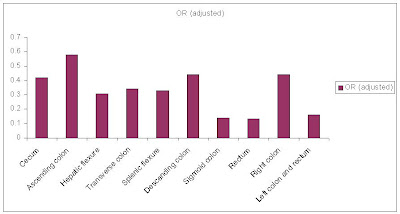
The findings of the study clearly show the benefit of colonoscopy in preventing colorectal carcinoma. It should be noted that colonoscopy by itself is not preventive, colonoscopy identifies the early adenomas which are removed. As development of colon cancer follows adenoma-carcinoma sequence, colonoscopy becomes protective by early detection and removal of adenomas. The study found that colonoscopy reduces the overall risk of colorectal carcinoma by 77%. This is substantial. The greatest risk reduction was seen in case of cancers located in the sigmoid colon (adjusted OR=0.14). The least risk reduction was for cancers located in the ascending colon. In general the risk reduction was more for left colon cancers than for right colon cancers.

efficacy of colonoscopy in reducing the risk of colorectal cancer
Adjusted odds ratio according to the site of colorectal cancer (less OR means more protection by colonoscopy).
The beneficial effect of colonoscopy did not vary with sex or with family history of colorectal carcinoma. The risk reducing effect of colonoscopy increased with increasing age in case of right sided colon cancer but such an effect was not observed in case of left sided colon cancers.

The study has few limitations like unaccounted confounders, possible differential rate of participation in the study. Also the authors did not account for the dietary habits of the participants. Increased intake of dietary fiber has a protective effect in development of colorectal cancer while intake of red meat and animal fat has a deleterious effect. It can be argued that BMI is a measure of the diet but I don’t think it can replace dietary history. This is one major point that I think the authors should have addressed.

ResearchBlogging.org
Brenner H, Chang-Claude J, Seiler CM, Rickert A, & Hoffmeister M (2011). Protection from colorectal cancer after colonoscopy: a population-based, case-control study. Annals of internal medicine, 154 (1), 22-30 PMID: 21200035

How aspirin might prevent colorectal cancer?

Posted: Sunday, January 2, 2011 | Posted by Debajyoti Datta | Labels: , 16 comments

I must confess that I am fascinated by the potential of aspirin in colorectal carcinoma prevention. It is worthwhile to look into this in some detail. Before we delve into the mechanism of colorectal carcinoma prevention by aspirin we must examine how colorectal carcinoma develops.


Colorectal carcinogenesis is characterized by a set of genetic alterations. The Fearon and Vogelstein model is a well accepted model explaining the sequence of colorectal cancer development. Their model is based on adenoma-carcinoma sequence. This suggests that colorectal carcinoma is preceded by adenomatous changes. Where adenomatous changes can not be identified, it is considered that the carcinoma directly arose from a dysplastic lesion. The development of colorectal carcinogenesis is taken to occur through through two distinct pathways –
  • APC/β-catenin pathway.
  • Microsatellite instability pathway.
In both the pathways there is step-wise accumulation of mutations but the genes involved and mutations occurring are different.

The APC/ β-catenin pathway is characterized by the loss of Adenomatous polyposis coli (APC) gene. The APC gene is located in the long arm of the 5th chromosome (5q21). APC is a dual-function tumor suppressor gene coding for a protein that binds to bundles of microtubules and promotes cell adhesion and migration. The level of β-catenin is also regulated by APC. β-catenin is a mediator in the Wnt/ β-catenin signaling pathway which plays a significant role in normal development of the intestinal epithelium. It is also involved in colorectal carcinoma development. Inactivated APC can be found in more than 80% cases of colorectal cases and 50% of the cases that don’t have APC mutation have β-catenin mutation.

β-catenin, a member of the cadherin based cell adhesive complex, acts as a transcription factor when translocated to the nucleus. In case of APC gene mutation, β-catenin accumulates in the cytoplasm and is subsequently translocated to the nucleus. When inside the nucleus it binds with a family of transcription factors called T-cell factor or lymphoid enhancer factor (TCF or LEF). β-catenin –TCF complex is considered to activate genes associated with regulation of cellular proliferation and apoptosis like c-MYC and CYCLIN D1. Thus mutation in APC gene leads to increased cellular proliferation and decreased cell adhesion.

Mutation of the oncogene K-RAS is thought to occur next. This is the most commonly activated oncogene seen in adenomas and colorectal carcinomas. Allelic loss at chromosome 18q21 occurs and it is suggested to be SMAD2 and SMAD4 which are involved in TGF β signaling. p53 gene mutations occur late in colorectal carcinogenesis and are seen in 70 to 80% cases of colon cancer. Increased telomerase activity has also been found in colorectal cancers.

The microsatellite instability pathway is characterized by genetic lesions in the DNA mismatch repair genes. These lesions are found in the HNPCC (Hereditary Nonpolyposis Colorectal cancer) syndrome and in 10 to 15% of sporadic cases. Inactivation of the DNA mismatch repair genes results in defective DNA repair. Any of the human mismatch repair genes hMSH2, hMLH1, MSH6, hPMS1, hPMS2 may be involved in HNPCC syndrome.

Mutations in mismatch repair genes cause alterations in microsatellites (fragments of repeat sequences in human genome that are prone to misalignment during DNA replication). This leads to microsatellite instability. Some microsatellites are located in the coding or promoter regions of genes like type II TGF – β receptor and BAX. TGF – β signaling is involved in inhibiting the growth of colonic epithelial cells and BAX genes cause apoptosis. Microsatellite instability thus leads to their nonfunctioning and colorectal carcinogenesis.

Now where does aspirin come in all these? Aspirin belongs to a group of drugs called NSAIDs (Non Steroidal Anti Inflammatory Drugs) that inhibit cyclooxygenase (COX) enzymes resulting in decreased prostaglandin synthesis. There are two isoforms of COX, COX-1 which is constitutively expressed and COX-2 which is inducible.

In colorectal carcinogenesis there is overexpression of COX-2 enzyme, there by giving aspirin the chance to bite. However the exact step where aspirin might act is unclear.

development pathway of colorectal cancer
Multistep progression of colon cancer and sites of NSAID action. From Postgrad Med J 2005;81:223-227 doi:10.1136/pgmj.2003.008227

Aspirin and other NSAIDs might also act through COX independent pathways to prevent colon cancer. High doses of aspirin have been shown to oppose the survival signaling pathway mediated by the transcription factor NF-kB. This is considered to occur through the inactivation of IkB kinase β. IkB kinase β is responsible for the activation of NF-kB cycle by phosphorylation of the inhibitory subunit of NF-kB. Hence aspirin inhibits the NF-kB pathway and interferes with cell survival.

pathway of action of aspirin in colorectal cancer prevention

Molecular mechanisms that mediate the effects of NSAIDs and anticancer drugs on survival and apoptosis in colon cancer cells. Schematic representation of cytokine, EGF-related growth factors and TRAIL ligand-dependent signal transduction pathways for survival and apoptosis. Stimulatory and inhibitory effects are indicated by arrows and bars, respectively. Abbreviations: MAPK=mitogen-activated protein kinase, MAPKK=mitogen-activated protein kinase kinase, JNK=jun kinase, IkB=inhibitor kinase B, NF-kB=nuclear factor kappa B, COX=cyclooxygenase, PI3K=phosphatidylinositol 3 kinase. From:Br J Cancer. 2003 March 24; 88(6): 803–807.
Synergistic effect of NSAIDs with conventional chemotherapeutic drugs has also been observed. The effect of NSAIDs in preventing neoangiogenesis is also important. Neoangiogenesis is a vital event in tumor growth and metastasis and tumor cells need adequate blood supply to derive nutrients. Prostaglandins are thought to be involved in angiogenesis by regulating proangiogenic factor synthesis like vascular endothelial growth factor (VEGF). Both COX-1 and COX-2 are considered to be involved in this.

Relative levels of Bcl-2 proteins regulate eukaryotic cell survival and apoptosis. SC-58125 and NS-398, COX-2 selective inhibitors, downregulates anti-apoptotic protein bcl-1 and sensitizes colorectal and proastate cancer cels to apoptosis. Aspirin upregulates bax and bak (proapoptotic proteins) and activates caspase 3 resulting in apoptosis. AKT, an anti-apoptotic protein kinase and the Fas-associated death domain has also been implicated in NSIAD induced apoptosis.

What remains to be seen is whether the advantages of aspirin will be sufficient considering the risk of GI adverse effects and how much benefit aspirin or any other NSAID provides. I want to remain hopeful.

Reference:
ResearchBlogging.org
Sangha, S. (2005). Non-steroidal anti-inflammatory drugs and colorectal cancer prevention Postgraduate Medical Journal, 81 (954), 223-227 DOI: 10.1136/pgmj.2003.008227
ResearchBlogging.org
Ricchi, P., Zarrilli, R., di Palma, A., & Acquaviva, A. (2003). Minireview: Nonsteroidal anti-inflammatory drugs in colorectal cancer: from prevention to therapy British Journal of Cancer, 88 (6), 803-807 DOI: 10.1038/sj.bjc.6600829
ResearchBlogging.org
KINZLER, K. (1996). Lessons from Hereditary Colorectal Cancer Cell, 87 (2), 159-170 DOI: 10.1016/S0092-8674(00)81333-1